
Serving East Central Illinois
Serving East Central Illinois
Champaign: 217-355-7494
Bourbonnais: 815-802-2090
Serving East Central Illinois
Serving East Central Illinois
Champaign: 217-355-7494
Bourbonnais: 815-802-2090
Diagnostic Technology
Diagnostic Technology
Retinal Exam
Your initial examination and testing will be comprehensive. Since we endeavor to complete all tests and make recommendations based on the results at the time of your exam, please allow several hours for your first visit (2-3 hours).
Your pupils will be dilated in order to perform our exam. Pupillary dilation will enlarge your pupils and make your eyes sensitive to bright light when you leave our office. For your safety, it is best to plan for someone to drive you from our office. The pupillary dilation will also prevent you from being able to focus, and reading may be difficult for several hours following our exam. You may wish to take this into consideration in your work schedule. These inconveniences are temporary but necessary for a complete exam.
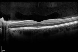
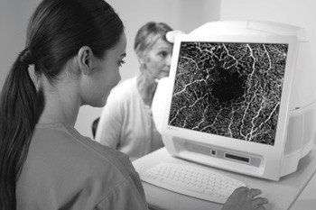
Optical Coherence Tomography (OCT)
Optical Coherence Tomography is a safe, non-invasive technique to obtain high resolution images of the retina. A beam of light is shined into the eye, and its reflectance is processed to create images of the retina. This technique does not involve any radiation (it’s not an x-ray or CT scan). State of the art Spectral Domain OCT images are crucial in diagnosing and managing many retinal conditions and glaucoma.

Retinal Angiography
In order to assess the health of blood vessels in the retina and choroid, a yellow dye called fluorescein is injected into a vein in the arm, and pictures are taken of the retina. These images are helpful in determining if the retinal vessels are healthy or damaged from a variety of retinal conditions such as diabetic retinopathy, macular degeneration, retinal vein occlusion, and others. Fluorescein dye can sometimes cause brief nausea, usually turns the urine and skin yellow-orange for several hours, and can rarely cause severe anaphylactic reactions.
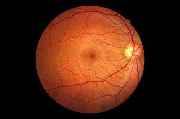
Fundus Photography
A special camera is used to obtain photographs of the optic nerve and retina. This allows us to document retinal pathology and, based on subsequent photographs, track its progress. The extent and severity of many retinal conditions can be monitored using fundus photography.
Fundus Photography
A special camera is used to obtain photographs of the optic nerve and retina. This allows us to document retinal pathology and, based on subsequent photographs, track its progress. The extent and severity of many retinal conditions can be monitored using fundus photography.
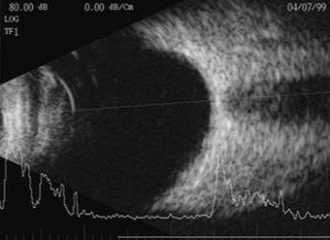
Ocular Ultrasound
B scan ultrasonography uses sound waves to generate images of the back of the eye. This is a painless, non-invasive technique, in which a probe is lightly placed on the eye. This is very helpful in assessing the vitreous, retina and choroid when a dense cataract or vitreous hemorrhage precludes a good view of the retina.
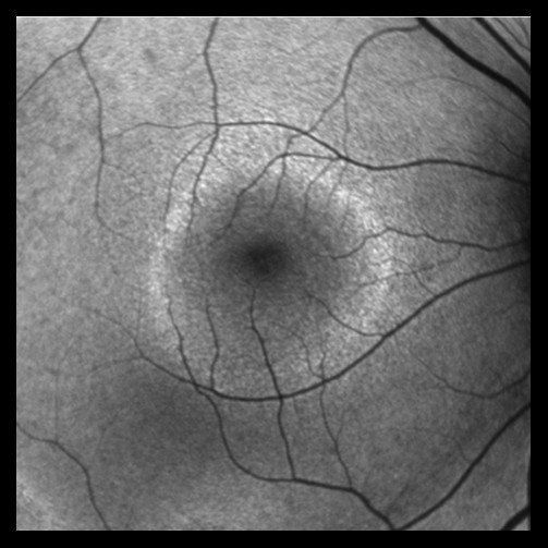
Autofluorescence imaging
A newer technique uses a blue-green wavelength and special filters to highlight the health of the retina and macula, and is based on assessing the presence or absence of certain molecules in the retinal pigment epithelium. Serial images provide evidence of disease progression over time. This is especially helpful in macular degeneration and plaquenil maculopathy.
OCTA:
Another newer non-invasive technology, OCTA (Angiography) uses advanced computer processing to determine blood flow in the retina and choroid, without the use of a dye. This can be especially helpful in highlighting areas of abnormal blood vessels in the retina and choroid.
OCTA:
Another newer non-invasive technology, OCTA (Angiography) uses advanced computer processing to determine blood flow in the retina and choroid, without the use of a dye. This can be especially helpful in highlighting areas of abnormal blood vessels in the retina and choroid.